Preclinical MRI scanners usually have between 1 and 4 independent receiver channels. However, there are many applications in which a higher number of receiving elements can be interesting, for example when willing to perform some of the following tasks:
- General anatomic localization imaging.
- Detail imaging of a region of interest.
- Imaging of second region of interest, smaller than or not connected to the previous one.
- Spectroscopy analysis on a small region, previously localized through imaging.
In order to achieve these objectives, it is necessary to change from one receiver coil configuration to another, choosing which physical elements of the receive array are active and connected to the scanner in each moment. This procedure allows to activate one imaging area or another without needing to physically move the animal over the coil, and without having to perform any new adjustmente.
Although this technique is very common in human MRI scanners, Neos Biotec is the only manufacturer offering it for preclinical MRI scanners.
Amongst other advantages, variable configuration arrays lead to a better use of the receiver channels of the MRI scanner, an improvement in SNR for each of the different imaging / spectroscopy regions of interest, and a more efficient work flow avoiding a lot of animal repositioning + adjustment tasks.
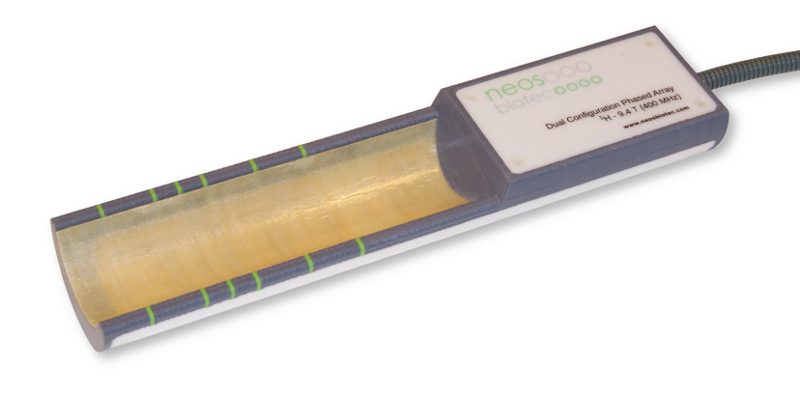
Example of a variable configuration phased array for mouse imaging (download datasheet):
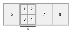
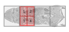
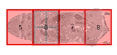
- Configurations: 2 (cardiac imaging / whole body imaging)
- Number of independent receiver channels in the MRI scanner: 4
- Number of array elements: 8 (7 physical elements + 1 virtual element, result of the quadrature combination of 4 physical elements)


